“I will also ask for an appropriation of an extra $100 million to launch an intensive campaign to find a cure for cancer, and I will ask later for whatever additional funds can effectively be used. The time has come in America when the same kind of concentrated effort that split the atom and took man to the moon should be turned toward conquering this dread disease. Let us make a total national commitment to achieve this goal.”
President Richard M. Nixon (State of the Union Address, 1971)
This year, 2011, is the fortieth anniversary of the War on Cancer. President Richard Nixon signed the National Cancer Act on December 23, 1971 inaugurating the “War on Cancer.” Since 1971 the War on Cancer has consumed an astonishing $200 billion, with the annual budget of the National Cancer Institute alone now at over $5 billion. This is comparable to the annual rate of expenditure of the Manhattan Project which invented the first atomic bombs and nuclear reactors between 1939 and 1945, with most of the work and expenditure between 1942 and July 16, 1945, the date of the first atomic bomb test known as Trinity, continued for forty years. The Manhattan Project consumed about $20 billion in 2011 dollars, about $2 billion in 1940’s dollars. The War on Cancer is roughly comparable to ten Manhattan Projects. The results have clearly been very disappointing. Since cancer is one of the leading causes of death, almost everyone would almost certainly like to see much more impressive results than achieved so far.
The War on Cancer was inspired in part by the spectacular success of the wartime Manhattan Project, the subsequent development of the hydrogen bomb (1945-1952), and the then (1971) recent spectacular success of the Apollo Program (1962-1969). This inspired not only the War on Cancer but many other “new Manhattan Projects” such as research into tokamaks and inertial confinement fusion devices for fusion power. Like the War on Cancer, most of these “new Manhattan Projects” have yielded disappointing results, certainly nothing on the scale of the Manhattan Project or the Apollo Program. As discussed in the previous article “The Manhattan Project Considered as a Fluke,” the Manhattan Project appears to have been a fluke, atypical of major inventions and discoveries especially in the success of the first full system tests: the Trinity test explosion on July 16, 1945 and the atomic bombings of Hiroshima and Nagasaki. Theoretical mathematical calculations and primitive numerical simulations seem to have been unusually successful in the case of the Manhattan Project compared to other breakthroughs. The Manhattan Project was probably quite unusual among major inventions and discoveries in other ways as well, but this is less clear. In terms of funding levels and the serious life and death nature of the goal, the War on Cancer is one of the closest analogs to the Manhattan Project among the many “new Manhattan Projects” of the last forty years.
With the widespread availability of extremely powerful computers, there are increasing attempts to apply mathematics and computational methods to biology and to cancer. There is a burgeoning field of “quantitative biology,” which includes its own section on the popular arxiv.org electronic preprint server. In many respects, this is an attempt to replicate the apparent success of theoretical mathematical calculations and early computer simulations in the Manhattan Project (1939-1945), the development of the hydrogen bomb (1945-1952), and the Apollo Program (1962-1969).
This article discusses the application of mathematics to the cure of cancer, the possible use of systems of smart drugs to perform simple mathematical calculations to identify and kill cancer cells, and presents a possible mechanism, developed by the author several years ago, to selectively destroy cells with abnormal numbers of chromosomes (aneuploidy), something common in many forms of cancer.
What does forty years of failure mean?
Such long periods of repeated failure are common in the history of scientific and technological breakthroughs. In most cases, this repeated failure has reflected either a lack of fundamental knowledge or an incorrect assumption or group of assumptions that was widely, even universally, held. While these two categories are not sharply defined and blur together, in general a lack of fundamental knowledge means that the state of knowledge was simply far too primitive to solve the problem. An example of this would be the failure of alchemists for thousands of years to transform base metals into gold or produce an elixir of life, the two major goals of both Western and Eastern alchemy. Today, we can with great difficulty and at great cost convert base metals into gold and the elixir of life remains a distant dream. It is clear in retrospect that the alchemical theory that metals were a composition of mercury and sulphur was grossly in error as were many other concepts of alchemy. Nonetheless, alchemists made many significant technological advances including methods for creating alloys similar to gold, gold and gold-colored coatings, and the discovery of a wide variety of useful materials. These successes, unappreciated today, probably gave the alchemists a false confidence in their theories and knowledge.
The blurriness of the two categories is illustrated by asking what might have happened if the alchemists had questioned and abandoned the mercury-sulphur theory which in various forms was widely held for many centuries. Loosely, the mercury-sulphur theory of metals postulated that metals were composed of mercury and sulphur in varying proportions; gold being mostly mercury, for example. This theory is frequently attributed to the Islamic alchemist Jabir ibn Hayyan (born in about 721 in Tus, Iran, died in about 815 in Kufa, Iraq) also known as Geber in Latin. Based on current knowledge, the alchemists would have had to have abandoned the mercury-sulphur theory and isolated a radioactive material such as uranium or invented batteries and other electrical technologies leading to particle accelerators to have had any hope of achieving their goal, both of which require performing very different experiments from the ones alchemists typically did. Batteries, in particular, could have been invented many centuries before they came into widespread use in the early nineteenth century.
In many cases, in retrospect, it is clear that this pronounced lack of progress in solving a scientific or technological problem was due to an assumption or group of assumptions that were incorrect and widely held. Indeed, often the assumption was something viewed as self-evident, something “everyone knows,” obvious, firmly established by extensive evidence, and so forth. Only in retrospect is it “obvious” that the assumption or assumptions were in error and not well supported by evidence, experience, or logic as most believed. Hence, one should ask whether some widely held, seemingly sensible belief or group of beliefs, supported by “overwhelming evidence” in modern scientific jargon, in biology is not, in fact, wrong.
In principle, the Internet and specific new technologies such as HTML or wiki’s should make it easier for researchers to collaborate and to list all assumptions, both stated and unstated, in a research field and their interdependencies, including links to all supporting raw data, experiments, and logical arguments: something like a Biology and Cancer Assumptions Wikipedia, but more rigorous than Wikipedia. Identifying and evaluating assumptions can be done much more systematically and thoroughly with hypertext, databases, and other Internet and computer software than was possible a few years ago using books, research papers, and conference presentations.
Questioning assumptions, especially assumptions “everyone knows,” is a social and political process. In modern Big Science, certain usually foundational assumptions are closely associated with high status individuals and institutions and are routinely presented with few qualifications or even as proven fact beyond any rational questioning to the public, business leaders, and policy makers: in Scientific American articles, PBS/Nova video programs, congressional testimony, private informal discussions where decisions are often actually made, and so forth.
In modern scientific research, there is pervasive rhetoric about “questioning assumptions” and “thinking outside the box,” but this usually does not apply to the foundational assumptions mentioned above. This rhetoric usually refers to subsidiary assumptions such as which protein to use in a biology lab experiment and similar non-threatening technical minutia. Indeed, many fields that have shown little or no practical results for decades, like cancer research, are periodically swept by fads and fashions in which subsidiary assumptions are replaced, modified, or added.
The discussion of mathematical approaches to curing cancer below generally assumes that modern biology has it right. A discussion of assumptions that might be in error or unorthodox biological concepts is mostly beyond the scope of this article, although the theory that abnormal numbers of chromosomes might play a more important role in cancer than generally thought is discussed. This general acceptance of current assumptions in biology is an important qualification that readers should keep in mind. At least historical experience with many “hard problems” in science and technology would suggest otherwise — that modern biology is “missing something” as physicists like to say.
Current Mathematical Approaches
There are a number of current attempts to apply mathematics to the cure or treatment of cancer. Quite a number are attempts to use differential equations to model the growth and spread of cancers and their response to various treatments and the immune system, fairly similar to the work of Dr. Swanson and Professor Levy described in detail below.
A well known example is Dr. Kristin Swanson’s work at the University of Washington:
We specialize in the mathematical modeling of pathological biosystems – specifically, primary brain tumors known as gliomas. We are currently working on several collaborative projects utilizing both clinical and experimental imaging techniques such as MRI and PET.
Our focus is on: 1) predicting patient-specific tumor growth, 2) seeking patient-specific markers of tumor progression, and, 3) identifying predictors of response to therapy in individual patients.
In plain English, this is an attempt to predict the growth and spread of certain brain tumors using a mathematical model, usually differential equations, in order to carefully target the spreading cancer with radiation or other methods.
Another well known example is the work of Professor Doron Levy at the University of Maryland, College Park. From Professor Levy’s web site:
Together with Peter Lee (Hematology, Stanford University) and Peter Kim (University of Utah) we have been working on combining new experimental data and mathematical models to develop new methods for treating leukemia patients. The type of leukmia that we have extensively studied is chronic myelogenous leukemia (CML).
Our research emphasizes the role of the immune system in the progression of the disease. By now it is known that most patients have an anti-leukemia immune response. It remains a mystery as of why this immune response is incapable of providing a sufficient response to the disease.
Our main work in this field was published in the June 2008 issue of PLOS Computational Biology. In this paper we proposed to vaccinating CML patients using their own blood in order to boost their anti-leukemia immune response. Using mathematical models we showed that the key issue is to time the cancer vaccine based on the dynamics of the immune response of the individual patient. A vaccination that is provided to early or too late in the process (i.e. after diagnosis and the initiation of drug-therapy) will have no noticeable effect. Our calculations suggest that such a procedure may ultimately be used to cure the disease. The work assumes that patients are treated with Gleevec (imatinib) starting from the diagnosis of the disease. A timed vaccine may allow them to stop the drug therapy.
There are many other attempts to apply similar mathematics to the cure or treatment of various cancers including work by Larry Norton at Memorial Sloan-Kettering Cancer Center, Lisette de Pillis at Harvey Mudd College, Vito Quaranta and Alexander “Sandy” Anderson at the Vanderbilt Integrative Cancer Biology Center and the University of Dundee in Scotland, Franziska Michor at Memorial Sloan-Kettering Cancer Center, Sofia Merajver at the University of Michigan, Paul Macklin at the University of Southern California, and many others. An Internet search for “Mathematics and Cancer” will turn up many matches of this type.
The author is not too optimistic about these approaches, although they certainly have merit. It seems that one still would really prefer something that could identify and either safely destroy or somehow render harmless the actual cancer cells. Can mathematics do this or assist in doing this?
Smart Systems of Drugs
The current prevailing theory of cancer is the oncogene or “cancer gene” theory. This is viewed as a proven fact by many molecular biologists. The current (2011) Director of the National Cancer Institute, Dr. Harold Varmus, shared the Nobel Prize in Medicine with J. Michael Bishop for early work on this theory.
Cancer is now said to be hundreds, even thousands of different diseases. While a medical doctor or pathologist may identify something as “breast cancer” or “skin cancer” or a similar general category, at a molecular and genetic level, “breast cancer” is actually many different diseases. It is thought that cancer is caused by the accumulation of many mutations of many different oncogenes and tumor suppressor genes that control complex networks of proteins that direct the growth, functioning, and differentiation of cells. In biology, differentiation refers to the process by which cells “differentiate” during growth into various specialized types of cells such as neurons in the brain, blood cells, and so forth with different specific properties and functions.
One type of breast cancer may have genes A,B,C, and D mutated while another has genes W, X, Y, and Z mutated. Not only this, but the cancers are thought to be continually mutating and evolving in the body, developing immunity to chemotherapy drugs for example. Thus, there does not seem to be a common molecular target that an anti-cancer drug can target in the way that penicillin or other antibiotics can kill a wide range of different bacteria, for example. As a result the latest favored concept in cancer research and treatment has been “personalized” targeted drugs. If one can determine that patient X has genes A, B, C, and D mutated, then in principle one can select or produce a drug specific to this particular type of cancer with A, B, C, and D mutated. This, of course, requires developing not one or a few anti-cancer drugs, but hundreds or even thousands of anti-cancer drugs possibly further personalized for each patient. See, for example, this recent article (“Targeted drugs: the future of cancer treatment?”, The Huffington Post, June 9, 2011).
Personalized cancer treatment has suffered a recent high profile black eye with the retraction of several papers from a research group at Duke University and a front page article in the Friday, July 8, 2011 New York Times (“How Bright Promise in Cancer Testing Fell Apart“, by Gina Kolata, The New York Times, July 8, 2011, page A1). Cancer research has long been characterized by a series of research and treatment fads, with one wonder drug or treatment after another heavily touted for a time, followed by disappointment, and then replaced by a new wonder drug: interferon, interleukin-2, and many others in the last forty years.
Nonetheless, medical doctors and pathologists going back to Hippocrates seem to have been able to identify a single disease as cancer long before modern genetic methods. It may be that there are system level features of cancer cells that do identify them as cancer cells. Traditional chemotherapy drugs were designed to kill dividing cells on the theory that cancer cells divide rapidly. However, healthy cells divide also and traditional chemotherapy has very limited benefits if at all. Only surgical removal of a tumor before it spreads — becomes metastatic in cancer jargon — appears to be able to cure cancer using the common sense definition of “cure”. While targeting cell division largely does not work, targeting other system level characteristics of cancer may work.
Many readers are probably familiar with the concept of nanotechnology and nanorobots, usually associated with K. Eric Drexler. One can envision tiny robots, nanorobots, that enter the blood stream, analyze each cell in turn, and selectively kill the cancer cells. The fabrication of such nanorobots is far beyond our current or near future technology. We are nowhere near implementing a computer central processing unit (CPU) or a robot at a molecular level. Even if we could, we do not know how to program a nanorobot to recognize a cancer cell and distinguish it from a normal healthy cell.
What we might be able to do, with great difficulty, is produce a small system of interacting drugs/molecules that perform some mathematical calculation in the cell and selectively kill cells identified as cancer cells or probable cancer cells while leaving the normal healthy cells alone. It is here that mathematics may be of use. To achieve success in the near future, the simpler the mathematics the better. Even engineering a single molecule such as genetically engineered insulin for diabetics is a daunting task at present. So a system of even a few molecules would be a substantial and difficult undertaking.
There are some current attempts to pursue this approach, notably the Cure Cancer Project associated with Dr. Arnold Glazier, Dr. Emil Frei and others. The Cure Cancer Project accepts that the current main assumptions of biology and cancer research are correct. The notion is to identify an unchanging property of cancer cells that can be targeted by a system of smart drugs. In specific terms, this seems to refer to targeting certain patterns among proteins that are thought to be associated with certain general properties, proliferation and invasivenes, of malignant cancer cells. Dr. Glazier has written a book and made some presentations on his ideas.
The author’s educated guess is that an approach based on a system of drugs, perhaps along the lines of the Cure Cancer Project, is the most likely approach to produce an effective cure or treatment for most common forms of cancer in the near future. (This discussion is for educational and informational purposes only and is not an endorsement of the Cure Cancer Project.) An important caveat is that such approaches generally assume that current biological assumptions and theories such as the somatic mutation theory of cancer, the oncogene theory (a specific instance of the somatic mutation theory) and the Central Dogma of Molecular Biology (DNA is the boss) are correct, making a cure for cancer “just engineering.” Next, this article presents a possible method to destroy cells with abnormal numbers of chromosomes, a condition known as aneuploidy which is common in many cancers.
The Selective Destruction of Cells with Abnormal Numbers of Chromosomes
One common characteristic of many cancers is an abnormal number of chromosomes, known as aneuploidy. This is often an excess number of chromosomes. A normal healthy human cell has forty-six (46) chromosomes. Cancer cells often have more than forty-six chromosomes. This was discovered long before the modern genetic era. One historical theory, now out of favor, is that the abnormal number of chromosomes causes cancer. This theory is usually credited to the German biologist Theodor Boveri. The most prominent modern advocate of the role of aneuploidy and chromosomes in cancer is the extremely controversial researcher Peter Duesberg who has published some articles on his theories in cancer research journals and a popular article in Scientific American in 2007 (“Chromosomal Chaos and Cancer”, Scientific American, May, 2007). A number of other researchers such as Angelika Amon at MIT have been investigating the role of chromosomes and aneuploidy in cancer in recent years; references are given below. The abnormal number of chromosomes or the other chromosomal anomalies often seen in a wide range of cancers may be a system-level characteristic of cancer that could be targeted despite the extreme variation in gene-level mutations (part-level characteristics of cancer).
Even though there are over one-million research papers on cancer, it is difficult to get a clear picture of the role of aneuploidy in cancer. Most modern cancer research is conducted within the framework of the oncogene theory and an implicit assumption that the way to cure or treat cancer is to target either a protein generated by a cancer gene or the gene directly. Chromosomal anomalies, both abnormal numbers of chromosomes and the rearrangements of chromosomes that are common in many cancers, are usually discussed as an aside to the putative cancer genes. This translocation of chromosome X mutated the key cancer gene ABC, or the duplication of chromosome X resulted in two copies of the key cancer gene ABC.
It could be that killing cancer cells with the wrong number of chromosomes would have no effect on the disease. It would simply result in a cancer with the correct number of chromosomes in the surviving cancer cells. It could slow the disease if the abnormal number of chromosomes is related to the malignancy of the cancer cells. In the best case, it might cure the disease, if the abnormal number of chromosomes is either the cause of cancer or essential in some way to the malignant characteristics of the cancer cells.
It may be possible to kill cells with an abnormal number of chromosomes using a system of five molecules: a harmless precursor A, a source catalyst S, a cell killer B, a drain catalyst D, and a neutralized cell killer C that the cell can safely digest or excrete.
The source catalyst S is inactive until it bonds to a numerical or quantitative feature on the chromosomes such as the telomeres at the ends of the chromosomes or the centromeres at the center. It becomes an active catalyst S* when it bonds to the chromosomes. Then the activated catalyst S* catalyzes the conversion of a harmless precursor A into a cell killer B. The activated catalyst S* has a maximum throughput. If the concentration of the precusor A is high enough in the cells, the catalyst S* will add the cell killer to the cell at a rate proportional to the number of chromosomes in the cell.
The cell killer B is relatively harmless in low concentrations. It needs to build up to a high level to kill the cell. So far, this will happen in all cells. However, if there is a drain catalyst D that bonds to a numerical feature in the cell that is the same in both normal cells and abnormal cells (cancer cells) and becomes an active drain catalyst D* that removes the cell killer B by converting it to the neutralized cell killer C, then the concentration of B can be engineered to rise to lethal levels only in cells with too many chromosomes.
A ==>S*==> B
B ==>D*==> C
This system of drugs is like a bathtub with several running faucets, one for each chromosome, and a single drain. If there are too many faucets, chromosomes, the water level, the concentration of the cell killer B, will rise and overflow the bathtub. If there are the right number, forty-six, or too few, less than forty-six, faucets, the drain can remove the water being added and the water level never rises. The water level remains almost zero; the concentration of the cell killer B is way too low to harm the cell.
One can kill cells with too few chromosomes (less than forty-six) by swapping the roles of the drain and the source. The drain catalyst bonds to the chromosomes. The source catalyst bonds to the constant numerical feature of the cells. Thus, if there are too few chromosomes, there are not enough activated drains to remove the cell killer B produced by the source catalyst. The bathtub has one big faucet and many small drains, one for each chromosome.
In principle, one could eliminate all cells with either too many or too few chromosomes by first treating the patient with a system of drugs that kills cells with too many chromosomes and then a system of drugs that kills cells with too few chromosomes. Cancer cells are frequently reported to have too many chromosomes, but sometimes too few is also reported.
A computational system of this type would now (2011) be easy to implement using mechanical components like the gears and springs used in traditional mechanical clocks, vacuum tubes and other traditional analog electronics components, or an integrated circuit. The problem is that as simple as such a computational system is, it is extremely challenging to implement using our current ability to engineer proteins and molecular biological systems in the cell.
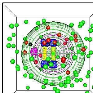
This video shows the build up of cell killer B in a cell with seven (7) chromosomes where the normal number of chromosomes is five (5). A smaller number of chromosomes than forty-six is used for demonstration purposes. The cell membrane is represented by a sphere which begins to distort when the cell killer concentration reaches the lethal level. The cell killer molecules are indicated by small green spheres that turn red when the lethal concentration is reached. The membrane disintegrates, killing the cell, and the cell killer disperses.
This graph shows the concentration of the cell killer B as a function of time:

Cell Killer Concentration
This video shows the lack of accumulation of cell killer B in a cell with five (5) chromosomes where the normal number of chromosomes is five (5).
This video shows the accumulation of cell killer B in a cell with three (3) chromosomes where the normal number of chromosomes is five (5). The sources and drains have been swapped as discussed above to kill cells with too few chromosomes.
This is the Octave script that simulates the bathtub mechanism and was used to make the videos above. This script runs successfully on a PC with Windows XP (Service Pack 2) using Octave 3.2.4. GNU Octave is a free, open-source high-level interpreted language, primarily intended for numerical computations that is mostly compatible with MATLAB.
cancer.m
function [result, command] = cancer(N_chromosomes, nsteps, bdisplay, lethal_level, cutoff, bTooFew, N_normal)
% [result, command] = cancer( [N_chromosomes, nsteps, bdisplay, lethal_level, cutoff, bTooFew, N_normal ])
%
% N_chromosomes - number of chromosomes in cell (default 50)
% nsteps - number of time steps to simulate
% bdisplay - boolean flag to display simulation (default false)
% lethal_level - concentration at which cells die (default 200)
% cutoff - number of time steps for cell death (default 10)
%          cell lives this many steps after the lethal concentration is reached
% bTooFew - simulate process to destroy cells with too few chromosomes
%           by using drains that attach to chromosomes and a source somewhere
%           in the cell. (default false)
% N_normal - number of chromosomes in a normal healthy cell (default 46)
%            sometimes set to small values such as 5 for demonstration or debugging
%
% Returns:
%      result -- time series of concentration of cell killer B
%      command -- ffmpeg command to create video of simulation
%
% Examples:
%
% [r, c] = cancer(7, 20, true, 10, 10, false, 5);  % kill cell with more than 5 chromosomes
% [r, c] = cancer(3, 20, true, 10, 10, true, 5);   % kill cell with less than 5 chromosomes
%
% Description:
%
% Basic Demonstration of Concept of Killing Cancer Cells by Chromosome Counting
% "Bathtub Mechanism" (conceived by John F. McGowan, Ph.D.)
% This is an Octave script (see https://www.gnu.org/software/octave/).
%
% N_chromosomes is the number of chromosomes in the cancer cell
%(46 is the number of chromosomes in a normal, healthy human cell).
%
% A (harmless poison precursor) =S=> B (poison) =D=> C (neutralized poison)
% A is harmless (ideally consumed orally or injected into bloodstream of patient).
% S is source on chromosome that catalyzes conversion of A to B
% B is toxic at concentration lethal_level (default 200)
% D is drain somewhere in cell that catalyzes conversion of B to C
% C is harmless.  Cell can safely digest or excrete C molecules.
%
% The sources on the chromosomes convert type A molecules to type B molecules.
% The drain (somewhere in the cell) converts type B molecules to type C molecules.
%
% Just like a bathtub drain, the drain in the cell has a limited throughput
% so if there are too many sources (too many chromosomes), the concentration of
% the toxic molecule B increases until it reaches the deadly lethal_level level and kills
% the cell.
%
% In most cancers, the cancer cells are aneuploid, they have the wrong number of
% chromosomes, usually too many.  If one can target the number of chromosomes in a cell
% one may be able to kill a wide range of cancers using a system of "smart drugs".
%
% In this example, the system of "smart drugs" consists of the poison precursor A, the
% sources (S) which bind to sites on the chromosomes, and the drain (D).  The sources only become active
% and able to catalyze the conversion of A to B when they bind to sites on the chromosomes.
%
% This does not require a complex Drexler style nanorobot or sophisticated pattern recognition
% to identify the cancer cells.
% The protein engineering is, of course, quite challenging, but not necessarily beyond present day
% or near future capabilities.
%
% Actual therapy would probably consist of infusing the source and drain molecules (S and D) into the patient.
% Once the sources and drains had attached to the cells (both healthy and cancerous), the precursor A
% would be introduced for a time until the cancer cells die.
%
% From "Can Math Cure Cancer?" by John F. McGowan, Ph.D., at The Math Blog
%
% Author: John F. McGowan, Ph.D.
% (C) 2011 by John F. McGowan, Ph.D.
% E-Mail: jmcgowan11@earthlink.net
%
%
concentration_a = 1.0;  % concentration of precursor A molecules
concentration_b = 0.0;  % concentration of cell killer B molecules
concentration_c = 0.0;  % concentration of neutralized C molecules

if nargin < 1
	N_chromosomes = 50;  % number of chromosomes in cell (46 is number in most normal, healthy human cells)
end

if nargin < 2
	nsteps = 100;  % number of time steps in the simulation
end

if nargin < 3
	bdisplay = false;  % display graphics of simulation
end

if nargin < 4
	lethal_level = 200;  % lethal concentration of B
end

if nargin < 5
	cutoff = 10; % number of time steps for cell death
end

if nargin < 6
	bTooFew = false;  % kill cells with too many chromosomes
end

if nargin < 7
	N_normal = 46;
end

N = N_normal; 46;  % normal number of chromosomes in a human cell (46)

printf("Normal Number of Chromosomes: %d\n", N);
fflush(stdout);

N_b = 0.0;  % number of molecules of cell killer b in cell
N_c = 0.0;  % number of neutralized molecules C in cell

volume = 1.0;  % volume of cell in arbitrary units

conc = [concentration_b];  % time series of concentration of cell killer B in cell

if bdisplay
	figure(1)  % figure 1 shows the B molecules in the cell
	[x_sphere,y_sphere,z_sphere] = sphere();  % get sphere to represent outer membrane of cell
end

delta_membrane = 0.5/cutoff;

nsteps_lethal = 0;  % number of steps since concentration reached lethal level

for i = 1:nsteps  % main simulation loop

	if bTooFew
		N_b += concentration_a * N;  % source S adds cell killer B to the cell
		delta_c = min(concentration_b, N_chromosomes);  % the drain D removes some cell killer B molecules (B =D=> C)
	else
		N_b += concentration_a * N_chromosomes;  % A =S=> B  (the sources on the chromosomes catalyze conversion of A to the cell killer B)
		delta_c = min(concentration_b, N);  % the drain D removes some cell killer B molecules (B =D=> C)
	end

	N_b = max(0, N_b - delta_c);  % drain converts B molecules to harmless C molecules
	N_c += delta_c;  % update number of neutralized C molecules in cell (cell can safely digest or excrete these molecules)
	concentration_c = N_c / volume;  % compute concentration of neutralized C molecules in cell
	concentration_b = N_b / volume;  % compute concentration of cell killer B molecules in cell

	if concentration_b >= lethal_level
		nsteps_lethal = nsteps_lethal + 1;
	end

	if nsteps_lethal > cutoff
	% cell dies so concentration of B starts to drop
	% membrane destroyed so molecule B disperses
	%
		if nsteps_lethal == cutoff
			printf('CELL DIES AT STEP %d', i);
			fflush(stdout);
		end
		N_b = floor(N_b / 2);
		concentration_b = N_b / volume;
	end

	conc = [conc concentration_b]; 	 % add new concentration of B to time series

	% display the B molecules
	% use ffmpeg (for example) to convert image sequence to video (e.g. mp4)
	% ffmpeg -r 10 -i cancer_50_chromosome_%03d.jpg cancer_50_chromo.mp4
	%
	if bdisplay
		if nsteps_lethal < cutoff  % membrane is destroyed for illustrative purposes (cell dies)
			mesh(x_sphere,y_sphere,z_sphere);
			% hard code limits of axes
%			xlim([-1.0 1.0]);
%			ylim([-1.0 1.0]);
%			zlim([-1.0 1.0]);
			hold on;
		else
			hold off
			% stop displaying the cell membrane after death
			clf(1);  % make sure figure is cleared
		end

		bpos = rand(N_b,3);
		x = bpos(:,1).*cos(2*pi*bpos(:,2)).*cos(2*pi*bpos(:,3));
		y = bpos(:,1).*sin(2*pi*bpos(:,2)).*cos(2*pi*bpos(:,3));
		z = bpos(:,1).*sin(2*pi*bpos(:,3));

		if concentration_b < lethal_level   % cell will die at concentration lethal_level
			scatter3(x, y, z, 12, [0 1 0]);  % use green color when concentration is below the lethal level
		else % lethal concentration level reached
			scatter3(x, y, z, 12, [1 0 0]);  % use red color when concentration is at or above the lethal level
			% distort membrane to indicate it is breaking down
			%
			x_sphere = (1.0 + delta_membrane) * x_sphere + delta_membrane*2*(rand(size(x_sphere))-0.5);
			y_sphere = (1.0 + delta_membrane) * y_sphere + delta_membrane*2*(rand(size(y_sphere))-0.5);
			z_sphere = (1.0 + delta_membrane) * z_sphere + delta_membrane*2*(rand(size(z_sphere))-0.5);
		end
		% hard code limits of axes
%		xlim([-1.0 1.0]);
%		ylim([-1.0 1.0]);
%		zlim([-1.0 1.0]);

		%view(60.0, 322.5);  % hard code view

		mytitle = sprintf('%3d B Molecules  Time Step: %03d', N_b, i);
		title(mytitle);
		frame_name = sprintf('cancer_%02d_chromosomes_%03d.jpg', N_chromosomes, i);
		print(frame_name);
		hold off;
	%	pause(1);  % optional pause of 1 second
	end % display
end % main simulation loop

result = conc;  % return time series of concentration of cell killer molecule B in cell
command = sprintf('ffmpeg -r 10 -i cancer_%02d_chromosomes_%%03d.jpg cancer_%02d.mp4', N_chromosomes, N_chromosomes);


% concentration of B increases without limit in cells with more than 46 chromosomes, reaches lethal level and kills cell.
% concentration of B stabilizes quickly at low level in cells with 46 or fewer chromosomes
%

if bdisplay
	figure(2)  % figure 2 shows the time series of the concentration of cell killer B molecules in the cell
	plot(conc);  % display time series
	mytitle = sprintf('Concentration of Cell Killer B (%d Chromosomes)', N_chromosomes);
	title(mytitle);
	fname_conc = sprintf('cancer_%02d_conc.jpg', N_chromosomes);
	printf("writing concentration time series to %s\n", fname_conc);
	fflush(stdout);
	print(fname_conc);
end

end % function cancer
In principle, the bathtub mechanism may be adapted to selectively kill cells with any abnormal quantity of any cellular feature, such as for example features on the exterior membrane of the cell (in which case the proteins must be trapped in the membrane surface). Thus, it may be adapted to targeting other numerical or quantitative abnormalities of malignant cancer cells than the number of chromosomes or the quantity of chromosomal features such as telomeres.
The presentation in this article is obviously simplified. For example, cells are multiplying and dividing. During cell division, a cell goes through a period where it has two sets of chromosomes before it splits into two cells. However, so long as the ratio of the sources to the drains is sufficiently constant integrated over time, the bathtub mechanism will still work and selectively destroy the dividing cells with too many or too few chromosomes.
This article does not discuss the specifics of how the five different molecules in the system might be implemented, most likely by modifying known molecules. This may be done in future articles or publications; the author does have some ideas how to implement the system in detail. Astute biologists may see how to implement some or all of the steps. It is likely that specialists from different sub-fields of molecular and cell biology will be needed to successfully implement the different parts of the system. Please feel free to contact the author if you have some good ideas.
Conclusion
Mathematics may be able to cure cancer through systems of smart drugs that perform a relatively simple calculation within a cell or on the membrane of the cell and kill or neutralize cells that are or probably are cancer cells. There are some attempts to develop a method along these lines. It does not seem to be the mainstream approach at the moment, which currently appears fascinated with personalized treatment, something of a legacy of the Human Genome Project and other gene-oriented research projects.
The author suspects that some common assumption or assumptions in biology and cancer research are in error or incomplete. This may be reflected not only in the evident lack of practical progress in cancer but also in the mostly failed attempts to cure or treat many other diseases in recent decades including most of the diseases currently identified as autoimmune disorders: Type I diabetes, multiple sclerosis (MS), systemic lupus, rheumatoid arthritis (RA), and so forth. Depending on what assumptions may be in error, all current approaches to curing cancer may be dead ends. If the unorthodox theory that abnormal numbers of chromosomes actually cause cancer is correct or a less drastic variant in which the abnormal number of chromosomes is essential to the malignant nature of cancer is correct, then a method that targets cells with an abnormal number of chromosomes, such as the method outlined in this article, may be successful. It is not clear which assumption or assumptions may be incorrect or incomplete; many other possibilities exist.
It may be possible to use modern Internet and computer software to collaboratively build an on-line database of biology and cancer research assumptions, both stated and unstated, their interdependencies, supporting data, and logical arguments to better identify and reevaluate current assumptions. Questioning assumptions, particularly those which "everyone knows," is a social and political process. It is not at all clear that we have the institutions and the mental outlook among researchers necessary to properly reevaluate assumptions in cancer or many other research fields. The rhetoric of "questioning assumptions" and "thinking outside the box" is easy to find in modern research. It is not "talking the talk," but rather "walking the walk" that is in question. On the other hand, most people and most cancer researchers would surely like to see major progress in the cure or treatment of the disease, since everyone faces a substantial risk of dying from cancer.
© 2011 John F. McGowan
About the Author
John F. McGowan, Ph.D. solves problems by developing complex algorithms that embody advanced mathematical and logical concepts, including video compression and speech recognition technologies. He has extensive experience developing software in C, C++, Visual Basic, Mathematica, MATLAB, and many other programming languages. He is probably best known for his AVI Overview, an Internet FAQ (Frequently Asked Questions) on the Microsoft AVI (Audio Video Interleave) file format. He has worked as a contractor at NASA Ames Research Center involved in the research and development of image and video processing algorithms and technology. He has published articles on the origin and evolution of life, the exploration of Mars (anticipating the discovery of methane on Mars), and cheap access to space. He has a Ph.D. in physics from the University of Illinois at Urbana-Champaign and a B.S. in physics from the California Institute of Technology (Caltech). He can be reached at jmcgowan11@earthlink.net.
Suggested Reading/References
Some Questions about Immortal Cell Lines
The Immortal Cell [Paperback]
Gerald B. Dermer (Author)
# Paperback: 212 pages
# Publisher: Avery (January 1, 1995)
# Language: English
# ISBN-10: 0895295822
# ISBN-13: 978-0895295828
# Shipping Weight: 12.8 ounces
A Conspiracy of Cells: One Woman's Immortal Legacy and the Medical Scandal It Caused [Hardcover]
Michael Gold (Author)
# Hardcover: 170 pages
# Publisher: State University of New York Press (January 1986)
# Language: English
# ISBN-10: 9780887060991
# ISBN-13: 978-0887060991
# ASIN: 0887060994
# Shipping Weight: 12.8 ounces
The Immortal Life of Henrietta Lacks [Paperback]
Rebecca Skloot (Author)
# Paperback: 400 pages
# Publisher: Broadway; Reprint edition (March 8, 2011)
# Language: English
# ISBN-10: 9781400052189
# ISBN-13: 978-1400052189
# ASIN: 1400052181
The Tissue Organizing Field Theory (TOFT) of Cancer
Wiley-Blackwell (2011, April 14). Controversial TOFT theory of cancer versus SMT model: Experts debate. ScienceDaily. Retrieved July 9, 2011, from https://www.sciencedaily.com /releases/2011/04/110415083217.htm
Ana M. Soto, Carlos Sonnenschein. The tissue organization field theory of cancer: A testable replacement for the somatic mutation theory. BioEssays, 2011; 33 (5): 332 DOI: 10.1002/bies.201100025
The Society of Cells: Cancer and Control of Cell Proliferation [Paperback]
Carlos Sonnenschein (Author), Anne Marie Soto (Author)
# Paperback: 154 pages
# Publisher: BIOS Scientific Publishers; 1 edition (January 1999)
# Language: English
# ISBN-10: 0387915834
# ISBN-13: 978-0387915838
Chromosomes, Aneuploidy, and Cancer
This is a collection of references, links, and abstracts of article on chromosomes, aneuploidy, and cancer.
Yuen, Karen Wing Yee(Oct 2010) Chromosome Instability (CIN), Aneuploidy and Cancer. In: eLS. John Wiley & Sons Ltd, Chichester. https://www.els.net [doi: 10.1002/9780470015902.a0022413]
--
Cancer Research
1.
Published OnlineFirst December 1, 2009; doi: 10.1158/0008-5472.CAN-09-2802 Cancer Res December 15, 2009 69; 9245
Cancer Stem Cells and Aneuploid Populations within Developing Tumors Are the Major Determinants of Tumor Dormancy
1. Anjali P. Kusumbe, and
2. Sharmila A. Bapat
https://cancerres.aacrjournals.org/content/69/24/9245.abstract
Human Molecular Genetics
Constitutional aneuploidy and cancer predisposition†
1. Ithamar Ganmore,
2. Gil Smooha and
3. Shai Izraeli*
+ Author Affiliations
1.
Sheba Cancer Research Center, Sheba Medical Center, Tel-Hashomer, Ramat Gan; Sackler Faculty of Medicine, Tel-Aviv University, Tel Aviv, Israel
1. *To whom correspondence should be addressed at: Pediatric Hemato-Oncology, Functional Genomics and Childhood Leukemia Research Section, Sheba Medical Center, Tel Hashomer 52621, Israel. Tel: +972 35303037; Fax: +972 45303031; Email: shai.izraeli@sheba.health.gov.il
* Received January 19, 2009.
* Accepted February 17, 2009.
* © The Author 2009. Published by Oxford University Press. All rights reserved. For Permissions,
1.
Hum. Mol. Genet. (2009) 18 (R1): R84-R93. doi: 10.1093/hmg/ddp084
https://hmg.oxfordjournals.org/content/18/R1/R84.short
--
Reference:
Identification of aneuploidy-selective antiproliferation compounds. Tang, Y-C., Williams, B.R., Siegel, J.J., and Amon, A. (2011). Cell. 144(4):499-512.
https://www.hfsp.org/frontier-science/awardees-articles/aneuploidy-new-anticancer-target
--
Mol Cell Biol. 2009 September; 29(17): 4766–4777.
Published online 2009 July 6. doi: 10.1128/MCB.00087-09.
PMCID: PMC2725711
Copyright © 2009, American Society for Microbiology
Loss of GATA6 Leads to Nuclear Deformation and Aneuploidy in Ovarian Cancer †
Callinice D. Capo-chichi,1,2 Kathy Q. Cai,2 Joseph R. Testa,2 Andrew K. Godwin,2 and Xiang-Xi Xu1,2*
Sylvester Comprehensive Cancer Center, Department of Medicine, and Department of Obstetrics and Gynecology, University of Miami School of Medicine, Miami, Florida 33136,1 Ovarian Cancer, Human Genetics, and Tumor Cell Biology Programs, Fox Chase Cancer Center, Philadelphia, Pennsylvania 191112
*Corresponding author. Mailing address: University of Miami School of Medicine, Rm. 417, Papanicolaou Building, 1550 NW 10th Ave. (M710), Miami, FL 33136. Phone: (305) 243-1750. Fax: (305) 243-5555. E-mail: xxu2@med.miami.edu
Received January 19, 2009; Revised February 23, 2009; Accepted June 25, 2009.
This article has been cited by other articles in PMC.
* Other Sections?
o Abstract
o MATERIALS AND METHODS
o RESULTS
o DISCUSSION
o Supplementary Material
o REFERENCES
Abstract
A prominent hallmark of most human cancer is aneuploidy, which is a result of the chromosomal instability of cancer cells and is thought to contribute to the initiation and progression of most carcinomas. The developmentally regulated GATA6 transcription factor is commonly lost in ovarian cancer, and the loss of its expression is closely associated with neoplastic transformation of the ovarian surface epithelium. In the present study, we found that reduction of GATA6 expression with small interfering RNA (siRNA) in human ovarian surface epithelial cells resulted in deformation of the nuclear envelope, failure of cytokinesis, and formation of polyploid and aneuploid cells. We further discovered that loss of the nuclear envelope protein emerin may mediate the consequences of GATA6 suppression. The nuclear phenotypes were reproduced by direct suppression of emerin with siRNA. Thus, we conclude that diminished expression of GATA6 leads to a compromised nuclear envelope that is causal for polyploidy and aneuploidy in ovarian tumorigenesis. The loss of emerin may be the basis of nuclear morphological deformation and subsequently the cause of aneuploidy in ovarian cancer cells.
https://ukpmc.ac.uk/articles/PMC2725711
--
[Proc Amer Assoc Cancer Res, Volume 45, 2004]
Cellular, Molecular, and Tumor Biology 89: Mouse Models of Prostate and Gastrointestinal Cancers
Abstract #4310
The role of the Bub1 gene in aneuploidy and cancer progression
Danaise V. Carrión, Marie Lia, Joerg Heyer, Patrick McDonald, Weijia Zhang, Kan Yang, Martin Lipkin, Ronan O’Hagan, Lynda Chin and Raju Kucherlapati
Harvard Medical School-Partners Center for Genetics and Genomics, Boston, MA, Albert Einstein College of Medicine, Bronx, NY, Strang Cancer Prevention Center, Rockefeller University, New York, NY, Dana-Farber Cancer Institute, Boston, MA
Human colorectal tumors can be classified on the basis of their genomic stability. Some CRC tumors show chromosomal instability (CIN) that is manifested by abnormal chromosome numbers while others show microsatellite instability. The cause for microsatellite instability is considered to result from mutations in DNA mismatch repair genes but the genetic basis for CIN is not well understood. We hypothesized that CIN might result from mutations in genes involved in the mitotic checkpoint. Bub1 is a gene involved in mitotic checkpoint. Mutations in Bub1 have been identified in CRC tumors, thus making this gene an excellent candidate to be involved in CIN and in colorectal cancer. To examine the role of Bub1 in carcinogenesis and chromosomal instability, we generated mice that carry a null mutation in this gene. Mice that are heterozygous for a null mutation in Bub1 are normal. When the heterozygotes were intercrossed, we failed obtain any homozygous offspring suggesting that Bub1 homozygosity leads to embryonic death. Bub1–/– embryos were detected at E3.5 but not at E8.5. We examined chromosome segregation in Bub1+/– and wild type (WT) ES cell lines. At different passages the Bub1+/– line showed a higher percentage of aneuploid cells. To confirm this observation FISH analysis was performed in blood cells of WT and Bub1+/– mice using probes for chromosomes 9 and 17. Modest chromosomal instability was observed in the Bub1+/– samples analyzed. Bub1+/– mice are fertile and susceptible to develop tumors very late in their lives. When Bub1+/– mice were bred to Apc1638N and Msh2 mutant mice, there was no difference in the incidence or multiplicity of tumors suggesting that the genomic instability provided by Bub1 heterozygosity is insufficient to significantly decrease the tumor latency or increase the incidence of tumors in the double mutants. Taken together, these results suggest that the Bub1 gene is essential for normal survival; and that heterozygosity of the gene leads to a mild chromosomal instability phenotype. This degree of chromosomal instability does not significantly affect the phenotype of Apc1638N and Msh2–/– mutant mice.
https://aacrmeetingabstracts.org/cgi/content/abstract/2004/1/994-d
--
Trends Cell Biol. 2005 May;15(5):241-50.
Aurora kinases, aneuploidy and cancer, a coincidence or a real link?
Giet R, Petretti C, Prigent C.
Source
CNRS UMR6061 Université de Rennes 1, Groupe Cycle Cellulaire, Equipe Labellisée LNCC, Université de Rennes 1, IFR140 GFAS, Faculté de Médecine, 2 Avenue du Pr Léon Bernard, CS 3417, Rennes cedex, France.
Abstract
As Aurora kinases are overexpressed in a large number of cancers, and ectopic expression of Aurora generates polyploid cells containing multiple centrosomes, it has been tempting to suggest that Aurora overexpression provokes genetic instability underlying the tumorigenesis. However, examination of the evidence suggests a more complex relationship. Overexpression of Aurora-A readily transforms rat-1 and NIH3T3 cells, but not primary cells, whereas overexpression of Aurora-B induces metastasis after implantation of tumors in nude mice. Why do polyploid cells containing abnormal centrosome numbers induced by Aurora not get eliminated at cell-cycle checkpoints? Does this phenotype determine the origin of cancer or does it only promote tumor progression? Would drugs against Aurora family members be of any help for cancer treatment? These and related questions are addressed in this review (which is part of the Chromosome Segregation and Aneuploidy series).
PMID:
15866028
[PubMed - indexed for MEDLINE]
https://www.ncbi.nlm.nih.gov/pubmed/15866028
--
Professor Angelika Amon (MIT)
The Cell Cycle and Cancer, Angelika Amon, June 7, 2006 (Video of Presentation to a General Audience)
Exploiting cancer cells' weaknesses
Team identifies potential drugs that enhance stress caused by too many chromosomes.
Anne Trafton, MIT News Office
https://web.mit.edu/newsoffice/2011/cancer-drugs-aneuploidy-0307.html
https://www.sciencedirect.com/science/article/pii/S0092867411000560
Volume 144, Issue 4, 18 February 2011, Pages 499-512
doi:10.1016/j.cell.2011.01.017 | How to Cite or Link Using DOI
Cited By in Scopus (7)
Permissions & Reprints
Article
Identification of Aneuploidy-Selective Antiproliferation Compounds
Purchase
$ 31.50
References and further reading may be available for this article. To view references and further reading you must purchase this article.
Yun-Chi Tang1, Bret R. Williams1, Jake J. Siegel1 and Angelika Amon1, Corresponding Author Contact Information, E-mail The Corresponding Author
1 David H. Koch Institute for Integrative Cancer Research and Howard Hughes Medical Institute, Massachusetts Institute of Technology, Cambridge, MA 02139, USA
Received 4 August 2010;
revised 22 November 2010;
accepted 17 January 2011.
Published online: February 10, 2011.
Available online 10 February 2011.
Referred to by: Targeting Aneuploidy for Cancer Therapy
Cell, Volume 144, Issue 4, 18 February 2011, Pages 465-466,
Eusebio Manchado, Marcos Malumbres
PDF (141 K) |
Summary
Aneuploidy, an incorrect chromosome number, is a hallmark of cancer. Compounds that cause lethality in aneuploid, but not euploid, cells could therefore provide new cancer therapies. We have identified the energy stress-inducing agent AICAR, the protein folding inhibitor 17-AAG, and the autophagy inhibitor chloroquine as exhibiting this property. AICAR induces p53-mediated apoptosis in primary mouse embryonic fibroblasts (MEFs) trisomic for chromosome 1, 13, 16, or 19. AICAR and 17-AAG, especially when combined, also show efficacy against aneuploid human cancer cell lines. Our results suggest that compounds that interfere with pathways that are essential for the survival of aneuploid cells could serve as a new treatment strategy against a broad spectrum of human tumors.
--
Modeling the Aneuploidy Control of Cancer
Li, Yao and Berg, Arthur and Wu, Louie R. and Wang, Zhong and Chen, Gang and Wu, Rongling (2010) Modeling the Aneuploidy Control of Cancer. BMC Cancer, 10 . Art. No. 346. ISSN 1471-2407 https://resolver.caltech.edu/CaltechAUTHORS:20100817-090307829
[img]
Preview
PDF - Published Version
Creative Commons Attribution.
442Kb
Use this Persistent URL to link to this item: https://resolver.caltech.edu/CaltechAUTHORS:20100817-090307829
Abstract
Background: Aneuploidy has long been recognized to be associated with cancer. A growing body of evidence suggests that tumorigenesis, the formation of new tumors, can be attributed to some extent to errors occurring at the mitotic checkpoint, a major cell cycle control mechanism that acts to prevent chromosome missegregation. However, so far no statistical model has been available quantify the role aneuploidy plays in determining cancer. Methods: We develop a statistical model for testing the association between aneuploidy loci and cancer risk in a genome-wide association study. The model incorporates quantitative genetic principles into a mixture-model framework in which various genetic effects, including additive, dominant, imprinting, and their interactions, are estimated by implementing the EM algorithm. Results: Under the new model, a series of hypotheses tests are formulated to explain the pattern of the genetic control of cancer through aneuploid loci. Simulation studies were performed to investigate the statistical behavior of the model. Conclusions: The model will provide a tool for estimating the effects of genetic loci on aneuploidy abnormality in genome-wide studies of cancer cells.
I enjoyed your article and hope to inspire my colleagues to join an interdisciplinary course where we read and study math, science, language, culture, etc. to keep the curing of diseases as one of the world’s best reasons for learning.
I had thought about something like this years ago, wondering about whether mathematics can have an impact on reducing cancer.
Biological “Logic Circuit” Destroys Cancer Cells
https://web.mit.edu/newsoffice/2011/cancer-detection-circuit-0902.html
Somewhat along the lines of what is discussed in “Can Mathematics Cure Cancer?” but it does not use a general characteristic of cancer cells such as abnormal numbers of chromosomes.
John
Respected sir
My self munish dadhwal from India thanks you straight from my heart for such a beautiful article on cancer.
I am a mathematics teacher in India teaching calculus for last seven years .but after reading your article i am so excited that i have published your article every where, in schools ,in my institute .sir my strong desire is to work on your project if you think i am capable to help. Three years before my FATHER dies because of kidney failure .only stupid method which is present is dialysis. I think if mathematics is able to help medical science then it is honor for us. Presently i am teaching calculus to high school students .i have done masters in mathematics
Regards Munish Dadhwal
Contact number 919872500605
John,
Thanks for the mention above. I was really thrilled when I realized you wrote the AVI guide awhile back–that was a very helpful resource when I was toying with EasyBMPtoAVI.
I’d be happy to answer any questions you might have on some of the current trends in mathematical cancer. (I’m personally mostly interested in developing techniques to calibrate current models to individual patient pathology, so that we can final validate/invadliate the models and dig at the underlying biology.)
All the very best — Paul
I’m not an intellectual but as I was doing my Highschool homework for Math I was thinking. Since the mathematical term e is seen in the world around us could cancer be possible connected to the natural log or sin e^x ? I have no clue but I thought there are no wrong questions or answers and I couldn’t find anything in my quick bing/google search about it being connected. I hope I did not waste your time for I sadly have to get back to doing homework. Keep up the fabulous works. 🙂